Science
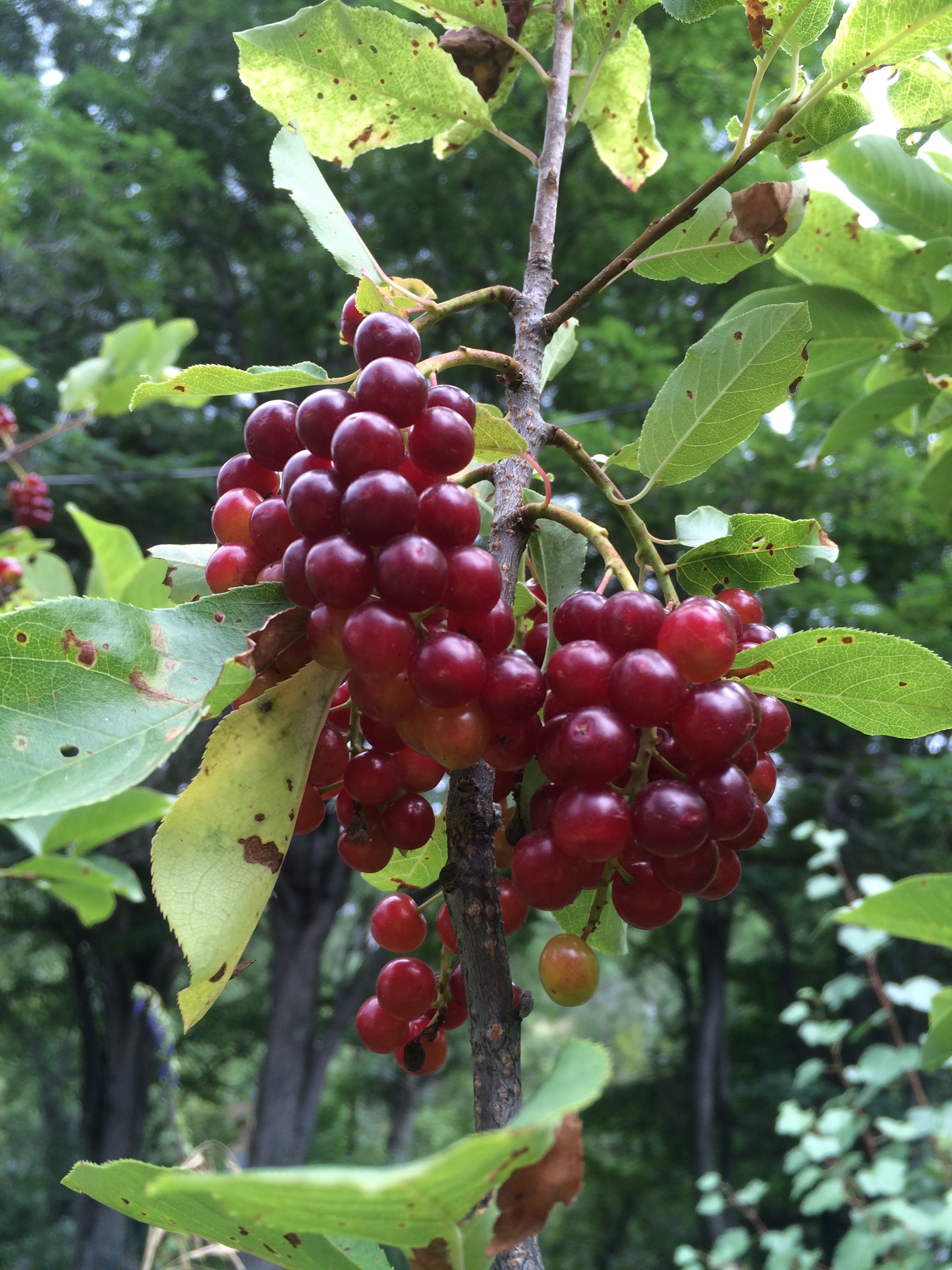
In the temperate zones of the Earth, late summer into autumn has been a time of celebration in many cultures. This is the time when all creatures breathe a sigh of relief as the hard work of growth slows. The cooler air transforms summer’s searing rays of sunshine into loving, golden warmth. Pregnant with sugar, fruits of flowering plants hang heavy from the branches and dapple the landscape in a mosaic of reds, blues and purples from anthocyanins and carotenoids. On the ground, combinations of lutein and zeaxanthin color the winter squashes of the Cucurbita family with the same oranges and yellows that are revealed as chlorophyll relinquishes its dominion over the foliage.
Colorful pigments that once acted as a beacon for pollinators in an array of colors and hormones[1] assume a new form that will serve as this year’s bridge of survival for numerous species of birds and mammals, including humans.
Over these precious few weeks, concentrated glucose and fructose flow in like the ocean tide. With them, the stomach’s master hormones of appetite flip flop. Ghrelin’s waxing and leptin’s waning[2] impose an ever-rising voracity of appetite that has driven successful survival of species over hundreds of millions of years. Inside the sweet goodness lurks even more treasures. Fresh omega-six oils from seeds and grains give a fresh boost to dwindling eicosanoids that are crucial for cell-to-cell communication. Vitamin E, selenium[3], vitamin C and phytonutrients stand like a levy to ensure the rising tide of inflammation doesn’t breach its banks.
In Traditional Chinese Medicine Theory, this time of year was considered the fifth season associated with the Earth element. Warmth, sunshine, water and Earth have been magically transformed by a billion tiny seeds into a form that passes life’s nourishment unto us. In the Jewish tradition, this season beckons the new year known as Rosh Hashanah.
“Blessed are you, sovereign of the Universe who brings forth bread to the Earth…who has kept us in life, has sustained us and brought us to this season.” Torah
Lurking deep within the cell, all the way down to the nuclear membrane, a sugar-laden surge of insulin nudges a sleeping Goddess from her torpor. 2.1 billion years ago[4],[5] some of the earliest fungi birthed this goddess and time kindly bequeathed her unto humans. In science she is known as SREBP or sterol regulatory elemental binding proteins. She is the one who, as if by magic, signals that transformation of sugar into a form that can be stored for later use as triglycerides[6] and fat[7]. Without her, most animals in the temperate and arctic zones are unlikely to survive even one winter.
Because of SREBP’s, every cell can make its own LDL cholesterol for membrane repair and vitamin D synthesis. However, without a way to supply basic antioxidants to the cell, LDL quickly oxidizes. This transformation from Dr. Jeckel to Mr. Hyde damages everything it touches[8] and is considered to be one of the driving forces of atherosclerosis7. In order to protect her inner world and ensure a constant supply of antioxidants, SREBP must ask for a little help from one of her cousins in the liver, SREBP-1. While most of the cells of the body settle for glucose as an energy source, the liver engages in a more refined taste for fructose. In fact, liver cells are the only ones that can use fructose and its effects are incendiary. Fructose drives rapid production of LDL cholesterol, fats and inflammation in the liver[9],[10]. This preference for fructose acts as a supply chain for the trillions of cells’ insatiable need for antioxidants during times like these. But without SREBP, these antioxidants are useless. She alone is the key master who permits passage of these antioxidants across the cell membrane. Under the dominion of SREBP, the LDL cholesterol receptor rises to the surface of the cell like a fish rising to feed. If it is lucky, LDL cholesterol will land in its mouth. Along for the ride, precious antioxidants like vitamins A, C, and E are granted access to the cell’s inner world[11].
As this season wanes, berries hang dried and scant on the branches. Insulin recedes as the sugar festival comes to a close. The Earth cools. SREBP breathes a deep sigh as her hard work comes to an end. As she falls into her winter nap, she brings many of the creatures of the Earth with her. Only one creature has successfully escaped the dominion of this goddess. Humans innovated to store carbohydrates externally. This consistent supply of sugar drives insulin to ensure that SREBP never sleeps. Her unrelenting state of slavery drives disorders like obesity[12],[13], fatty liver[14], insulin resistance[15] and atherosclerosis[16], [17]. Perhaps this goddess would argue that these are not diseases at all but are phenotypes brought on by depriving her of a proper rest.
[1] Cutler A.J., Krochko J.E. Formation and breakdown of ABA. Trends Plant. Sci. 1999;4:472–478. doi: 10.1016/S1360-1385(99)01497-1
[2] Teff KL, Elliott, SS, Tschop M, Kieffer TJ, Rader D., Heiman M., Townsend RR., Keim NL, D’Alesso D, Havel Dietary fructose reduces circulating insulin and leptin, attenuates postprandial suppression of ghrelin, and increases triglycerides in women. PJ J Clin Endocrinol Metab. 2004 Jun;89(6):2963-72
[3] Giacomo dugo, Lara La Pera, Donatella Pollicino, marello Saitta. Determination of Selenium Content in Different Types of Seed Oils by Cathodic Stripping Potentiometry (CSP) J. Agric. Food Chem., 2003, 51 (19), pp 5598–5601
[4] Timothy F. Osborne, Peter J. Espenshade Evolutionary Conservation and Adaptation in the Mechanism that Regulates SREBP Action: What a Long Strange tRIP It’s Been. Genes & Dev. 2009. 23: 2578-2591, doi:10.1101/gad.1854309
[5] V Laudet Evolution of the Nuclear Receptor Superfamily: Early Diversification from an Ancestral Orphan Receptor. Journal of Molecular Endocrinology Dec. 1, 1997. 19 2-7-226
[6] Colleen K. Nye Glyceroneogenesis Is the Dominant Pathway for Triglyceride Glycerol Synthesis in Vivo in the Rat The Journal of Biological Chemistry, 283, 27565-27574. October 10, 2008
[7] Hitoshi Shimano, SREBPs: physiology and pathophysiology of the SREBP family. The FEBS Journal 2009 276:3 616-621
[8] Low Density Lipoprotein Can Cause Death of Islet β-Cells by Its Cellular Uptake and Oxidative Modification Miriam Cnop, Jean Claude Hannaert, Annick Y. Grupping, and Daniel G. Pipeleers Endocrinology 2002 143:9 , 3449-3453 http://dx.doi.org/10.1210/en.2002-220273
[9] Zhang C, Chen X, Zhu RM, Zhang Y, Tu T, Wang H., Zhao H, Zhao M, Ji YL, Chen YH, Meng XH, Wei W, Xu DX. “Endoplasmic reticulum stress is involved in hepatic SREBP-1c activation and lipid accumulation in fructose-fed mice.” 2012 Aug 3;212(3):229-40. doi: 10.1016/j.toxlet.2012.06.002. Epub 2012 Jun 12.
“ER stress contributes, at least in part, to hepatic SREBP-1c activation and lipid accumulation in fructose-evoked NAFLD.”
[10] Koo HY, Miyashita M, Cho BH, Nakamura MT. Replacing dietary glucose with fructose increases ChREBP activity and SREBP-1 protein in rat liver nucleus. 2009 Dec 11;390(2):285-9. doi: 10.1016/j.bbrc.2009.09.109. Epub 2009 Sep 30.
“Nuclear SREBP-1 was 2.2 times higher in fructose-fed rats than glucose-fed rats.”
[11] Maret G Traber, Herbert J Kayden “Vitamin E is Delivered to Cells via the High Affinity Receptor for Low-Density Lipoprotein” The American Journal of Clinical Nutrition 40: October 1984, pp 747-51.
[12] Hitoshi Shimano, SREBPs: physiology and pathophysiology of the SREBP family. The FEBS Journal 2009 276:3 616-621
[13] Hitoshi Shimano, SREBPs: physiology and pathophysiology of the SREBP family. The FEBS Journal 2009 276:3 616-621
[14] Moon YA, Liang G, Xie X, Frank-Kamenetsky M, Fitzgerald K, Koteliansky V, Brown MS, Goldstein JL, Horton JD. The Scap/SREBP pathway is essential for developing diabetic fatty liver and carbohydrate-induced hypertriglyceridemia in animals. Cell Metab. 2012 Feb 8;15(2):240-6
[15] Iichiro Shimomura, Robert E. Hammer, James A. Richardson, Shinji Ikemoto, Yuriy Bashmakov, Joseph L. Goldstein,Michael S. Brown
Insulin resistance and diabetes mellitus in transgenic mice expressing nuclear SREBP-1c in adipose tissue: model for congenital generalized lipodystrophy. Genes Dev. 1998 October 15; 12(20): 3182–3194.
[16] Karasawa T, Takahashi A, Saito R, Sekiya M, Igarashi M, Iwasaki H, Miyahara S, Koyasu S, Nakagawa Y, Ishii K, Matsuzaka T, Kobayashi K, Yahagi N, Takekoshi K, Sone H, Yatoh S, Suzuki H, Yamada N, Shimano H. Sterol regulatory element-binding protein-1 determines plasma remnant lipoproteins and accelerates atherosclerosis in low-density lipoprotein receptor-deficient mice. Arterioscler Thromb Vasc Biol. 2011 Aug;31(8):1788-95.
[17] Kurtak, K. Dietary and Nutritional Manipulation of the Nuclear Transcription Factors, PPAR’s and SREBP’s, as a Tool for Reversing the Primary Diseases of Premature Death and Aging. Rejuvenation Research 17-2. April 2014. P 140-44.
A new study on the “bad luck” of cancer is a wonderful contribution to science but is being severely misinterpreted by both science writers and the media.
This study[1] is the first of its kind to accurately quantify the probability of the development of a cancer cell in any given tissue over a lifetime. It supports other hypotheses[2] stating that increased frequency of cell division, which is also a hallmark of cellular aging, leads to increased risk of cancer. However, it is not representative of a cancer cell’s ultimate destiny. New cancer cells form in our bodies everyday and our immune system destroys them. We’ve known for 20+ years that tissues that are prone to faster cell division and turnover, like colon and skin, have a higher probability of developing cancer cells. This is why inflammation is strongly associated with the development of cancer. Inflammation from immune activity causes rapid damage and therefore places a high demand on the affected tissue for renewal by cell division. Thus, a higher frequency cell division results in a higher statistical probability that mutations will occur, cancer cells will develop and one of them might escape under the immune system’s radar. An example; we know that cigarette smoking leads to a much higher risk of developing lung cancer. There are two parts to this. One is the simple carcinogenicity of some of the chemicals in cigarette smoke. However, a much larger role is played by the fact that the body responds to cigarette smoke by launching an immune response that leads to increased inflammation, increased cellular replacement, impaired cellular death and diminished tissue cleanup[3],[4].
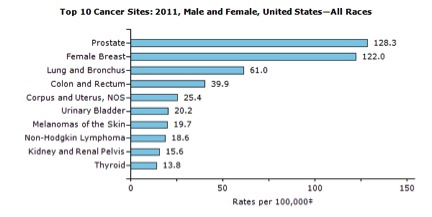
Most importantly, the cancer risk study excludes any statistics on breast and prostate cancers. Perhaps these were intentionally excluded from the study as edge cases since their occurrence in the general population is 300% higher than any of the cancers included in the study. Because these tissues are hormone-sensitive, they are highly susceptible to influence by external factors. Hundreds of studies have established their extreme vulnerability to chemicals that mimic estrogen and stimulate rapid growth and cell division. This parallels the theme of this recent study suggesting that faster cell division leads to a high probability of mutations and cancer cell development. Including these two types of cancer in the study’s statistics could increase their external influence factor by as much as 20%.
Although there are some correlations it is important to note that the probabilities in the study do not evenly parallel rates of cancer incidence in the US. For example the study shows, stem cell divisions in colorectal cells as being significantly higher but than in lung but cancer statistics show that the incidence of these cancers is flip flopped.
While this information is invaluable for quantifying cancer cell development, it is missing significant aspects of our basis of cancer knowledge and statistics and by no means establishes final numbers or parameters for cancer risk or its influence by external factors.
[1] Cristian Tomasetti, Bert Vogelstein. Variation in Cancer Risk Among Tissues can be Exlapined by the Number of Stem Cell Divisions. Science 2 January 2015 Vol. 347 no. 6217 pp. 78-81. DOI: 10.1126/science.1260825
[2] Steve Horvath. DNA Methylation Age of Human Tissue and Cell Types. Genome Biology 2013 12:R115 doi:10.1186/gb-2013-14-10-r115
[3] Naotaka Noda, Koichiro Matsumoto, Soturu Fukuyama, Yukari Asia, Hiroko Kitajima, Nanae Seki, Yuko Matsunaga, Keiko Kan-o, Atsushi Moriwaki, Konosuke Morimoto, Hiromasa Inoue and Yoichi Nakanishi. Cigarette Smoke Impairs Phagocytosis of Apoptotic Neutrophils by Alveolar Macrophages Via Inhibition of Histone Deacetylase/Rac/CD9 Pathways. Int. Immunol. (2013) 25 (11) 643-650. doi: 10.1093/intimm/dxt033
[4] Susan JM Hoonhorst, Wim Timens, Leo Koenderman, Adele T Lo Tam Loi, Jan-Willem J Lammers, H Marike Boezen, Antoon JM van Ossterhout, Dirkje S Postma, Nick HT ten Hacken. Increaded Activation of Blook Neutrophils After Cigarette Smoking in Young Individuals Susceptible to COPD. Respiratory Research 2014 15:121 doi:10.1186/s12931-014-0121-2
Hello to all of you wonderful people who have taken time out this precious life to read my blog. I had to check out for a while due to some personal tragedy. As some ancient cultures say ” I have been waiting for my soul to catch up with the rest of me” and I am FINALLY starting to write again. Upcoming subjects include:
-Evolution Dictates a Contrarian Approach to the Emergence and Spread of Human Pathogens
-Misinterpretation of Methylation. Why Folic Acid Doesn’t Cause Cancer
-A Conventional Interpretation of the Four Levels of Disease in Traditional Chinese Medicine
-A Conventional Interpretation of Digestion in Traditional Chinese Medicine
-Why We Need to Introduce a New School of Research if Science it Going to Successfully Evolve
I look forward to more great discussions and comments.
Sincere thanks to each of you!
Karen